AmnioGraft®
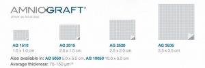
AmnioGraft is a biologic ocular transplantation graft used by eye surgeons around the world as an adjunct therapy to treat ocular surface indications such as corneal ulcers, pterygium, mechanical dry eye (also known as conjunctivochalasis), excision of tumors, chemical burns, and Stevens-Johnson Syndrome. AmnioGraft serves as a tissue replacement by delivering the unique therapeutic actions of cryopreserved amniotic membrane.
Expedite Recovery, Promote Regenerative Healing
AmnioGraft expedites patient recovery by reducing inflammation and scarring, promoting regenerative healing, and achieving a superior cosmetic outcome. This is particularly beneficial to many ocular surface diseases and conditions to ameliorate pain, vision loss, and in some cases an undesirable ocular cosmetic appearance.
The Healing Process
AmnioGraft is durable and easy to use, and provides lasting cosmetic results. After surgery, full healing is typically complete in two to three weeks.
What is amniotic membrane tissue?
Amniotic membrane is part of the placenta. It is the tissue closest to the baby throughout development in the womb. The biological properties of amniotic membrane protects the baby from any harm and its natural therapeutic actions helps the baby develop.
The placentas used to prepare AmnioGraft are donated by consenting mothers after cesarean section births. Mothers that donate are fully informed, have healthy lifestyles, and are tested against infectious diseases prior to donation.

Cutting-edge technology at the forefront of BioTissue!
Cutting-edge technology at the forefront of BioTissue!

Download AmnioGraft Product Information
Surgical Procedures
AmnioGraft improves surgical outcomes for a variety of surgeries including Mechanical Dry Eye (also known as Conjunctivochalasis), and Pterygium.
Tissue Tuck Procedure for Pterygium
Pterygium is a benign vascular pink tissue that can grow from the conjunctiva onto the cornea. If it grows into the line of vision, over the pupillary aperture, it can interfere with vision.
Pterygium is most commonly found to originate from the inner, nasal, surface of the eye and extend toward the pupil. They can also occur on the temporal side of the eye. Pterygium is believed to be caused by long-term and repetitive exposure to UV rays from sunlight. Therefore, Pterygium is more common in countries and states closer to the equator.
With Pterygium, the primary goals are to achieve no recurrence, fast recovery, and excellent cosmetic outcomes. The Tissue Tuck procedure leverages the physical and biological properties of AmnioGraft to make Pterygium surgery quick and easy to perform and creates two barriers to recurrence and the results are as follows:
- Faster recovery
- Minimized post-surgical inflammation
- Very low recurrence
- Reduced surgical time
- Superior cosmetic outcome in as little as 1 week
Listen to the Ophthalmology Management podcast below from Neel R. Desai, M.D., to learn more about the TissueTuck Technique, a new surgical approach to Pterygium surgery for low recurrence, better cosmesis, & shorter OR time. Find out:
1. How to perform premium pterygium surgery efficiently using cryopreserved amniotic membrane
2. How can you provide Wow Moments which lead to happy and loyal patients in your practice
3. How adopting the TissueTuck Technique helps you improve practice economics
Reservoir Restoration Procedure for Mechanical Dry Eye (Conjunctivochalasis)
Mechanical Dry Eye, also known as Conjunctivochalasis (CCh), is loosened, shortened and wrinkled conjunctiva, which interferes with the tear meniscus and diminishes the fornix or tear reservoir. This is caused by the underlying Tenon’s capsule being degenerated or dissolved by matrix metalloproteinases (MMPs). This wrinkled tissue occupies the tear reservoir and prevents the eye from holding tears, causing pain, tearing and irritation that feels similar to dry eye symptoms. Often, some of the wrinkled conjunctival tissue can disrupt the tear meniscus, at the edge of the eyelids. This tissue acts as the tip of an iceberg, which reflects only a portion of the underlying condition. When you blink, this wrinkled conjunctiva can rub against the eye, causing further irritation and redness to an already inflamed and dry eye.
Listen to the Ophthalmology Management podcast below from Dr. Douglas Devries, O.D., and Dr. Curtis Manning, M.D., of Eye Care Associates of Nevada on how they partner to diagnose and treat MDE to restore the patient’s anatomical tear reservoir, which addresses the underlying cause of mechanically induced dry eye and helps support:
· Efficient replacement of the degenerated Tenon’s fascia and excised conjunctiva
· Restoration of the tear reservoir to a healthy state
· Return of tear flow from fornix to tear meniscus
Mechanical Dry Eye (MDE), also known as Conjunctivochalasis, represents one of the most common age-related eye diseases and is characterized by the presence of redundant folds of the conjunctiva that typically are detected between the eyeball and the eyelids. It is commonly found along the lower lid margin and mechanically interferes with the normal distribution of tears giving rise to an unstable tear film (dry eye) and delayed tear clearance (epiphora). The differences between CCh-induced dry eye and aqueous tear deficiency (ATD) are summarized in the table below.
| DISTINGUISHING FEATURE | ATD DRY EYE | MECHANICAL DRY EYE |
|---|---|---|
| Diurnal Variation | Worse in PM | Same throughout the day |
| Worst gaze | Up gaze | Down gaze |
| Effect of vigorous blinking | Symptom improved | Symptom worsened |
| Recurrent subconjunctival hemorrhage | Infrequent | Frequent |
| Fluorescein Staining Pattern | Low tear meniscus without interruption | Tear meniscus interruption or obliteration |
| Tear Clearance | Normal/Delayed | Frequently delayed |
| Rose Bengal Staining | Exposure zone | Non-exposure zone |
| Effect of Punctal Occlusion | Symptom improved | Symptom worsened |
For asymptomatic MDE, no treatment is needed, and patients may be given tear substitutes, lubricants, corticosteroids or antihistamine drops. For MDE patients who do not respond to medical treatment, the Reservoir Restoration Procedure can resolve interference with the tear meniscus and using cryopreserved amniotic membrane will restore the tear reservoir to its normal state and expedite a patients’ recovery.
Other indications include:
- Corneal defects
- High-risk trabeculectomies
- Leaking glaucoma blebs
- Chemical burns
- Stevens-Johnson Syndrome
- Strabismus
- Removal of tumors
- And many other procedures…
Discover MDE
All you see is the tip of the iceberg.
AmnioGraft addresses the root cause of your dry eye, with most patients experiencing a fast recovery and a significant reduction in inflammation.
How To Use
How To Use

AmnioGraft Handling
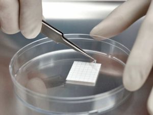
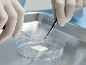
The Company’s unique cryopreservation method retains the tissue’s natural tensile strength, which makes this naturally strong tissue easy to handle and suture glue.AmnioGraft is supplied in an easy to use dual peel pouch. Use sterile smooth forceps or gloves to remove the inner pouch containing the tissue. The clear inner pouch may be introduced to the sterile field. Using sterile scissors, cut below the sealed line of the inner pouch and remove AmnioGraft using smooth sterile forceps. Once retrieved from the sterile, clear inner pouch, AmnioGraft can be easily removed from its carrier paper using a dry surgical sponge or 0.12 forceps.
The tissue is ready for transplantation immediately after removing it from the carrier paper without the need for rehydration.
AmnioGraft is always manufactured with the stromal side of the tissue attached to the carrier paper. It is easy to determine the orientation of AmnioGraft after it has been removed from its carrier paper using a dry surgical sponge. The dry sponge will stick to the stromal side, but it will not stick to the basement membrane side.
For packaging and storage info, click here.

REQUEST FOR MORE INFORMATION
Learn more about how AmnioGraft helps create a healing environment that is conducive for faster and more effective healing of the ocular surface, less scarring and inflammation, and improved clinical outcomes. Fill out the form below and a representative will contact you shortly.
