Neurotrophic Keratitis

Are you checking corneal sensitivity on your SPK patients?

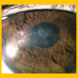
According to the Mackie classification, stage 1 Neurotrophic keratitis (NK) begins as early as Punctate epithelial keratitis (PEK).12

Discovering and Treating the Underlying Cause

Neurotrophic Keratitis (NK) is a corneal degenerative disease characterized by a reduction of corneal sensitivity.1 In NK, corneal innervation by the trigeminal nerve is impaired.1

This can lead to corneal epithelial breakdown, impairment of healing, and development of corneal ulceration, melting, and perforation.2 NK abolishes both tearing and blinking reflexes, thus causing the most severe forms of Dry Eye Disease (DED).3

Early intervention is critical in the treatment of NK.

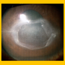
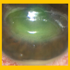
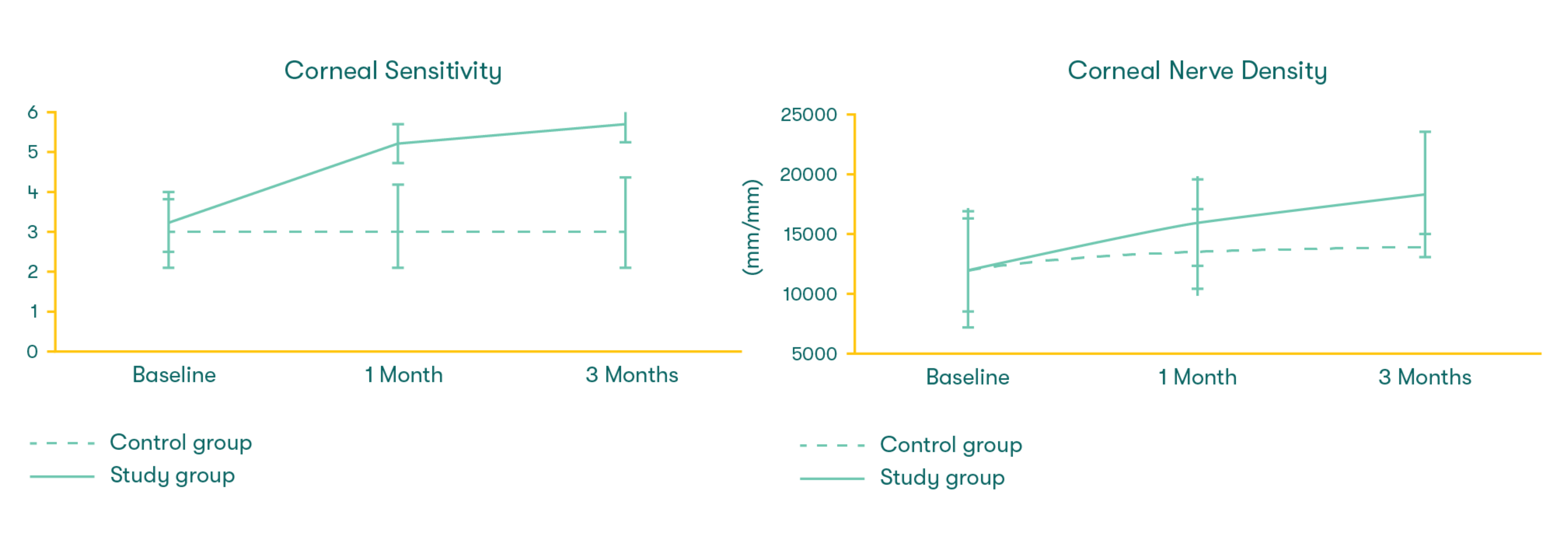
In a recent study by Dr. Thomas John, Prokera has been found to improve corneal nerve sensitivity and density.7 Early treatment is imperative to prevent further epithelial damage and potential vision loss 4-6. NK is a progressive condition with 3 stages of increasing severity (based on the Mackie classification):6,7

01

Punctate Epithelial Keratitis (PEK)

02

Persistent Epithelial Defect (PED)

03

Corneal Ulcer

Did you know the American Academy of Ophthalmology has recognized Prokera® as a stage 1 treatment option1?

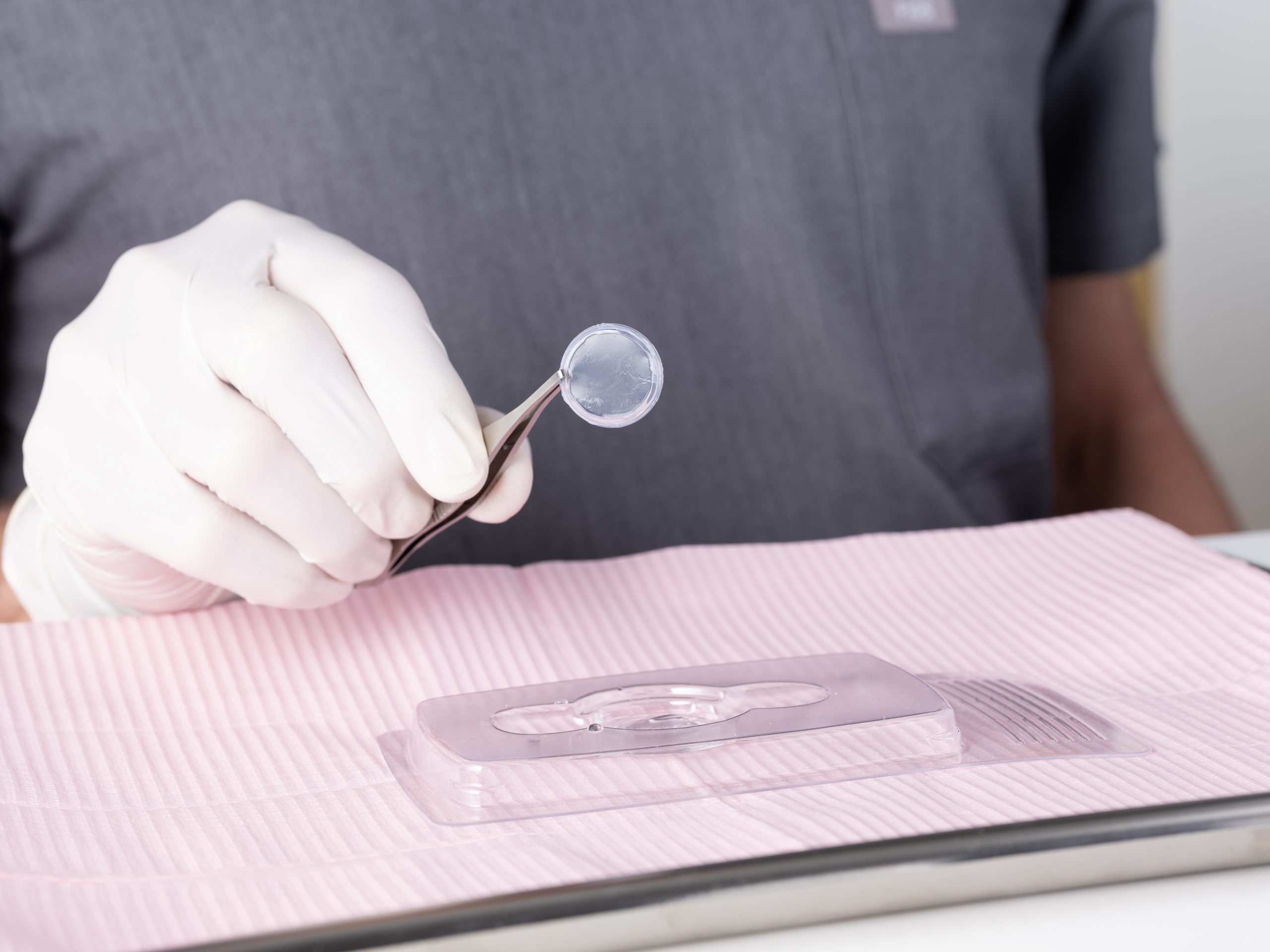
Prokera cryopreserved amniotic membrane helps regenerate corneal nerves as it accelerates the recovery of corneal surface health.7 Prokera has been shown to significantly reduce the signs and symptoms of DED as stated in Thomas John’s study.7 Prokera has also been used successfully to treat corneal defects and ulcers caused by NK, which were non-responsive to prior treatment as reported in other prospective studies.9-11
Download Prokera Product Information

To learn more about Prokera, fill out the details below:
By clicking the button below, I agree to receive marketing communications from BioTissue I am aware that I can unsubscribe at any time.
This field is for validation purposes and should be left unchanged.

How can I test for Neurotrophic keratitis?

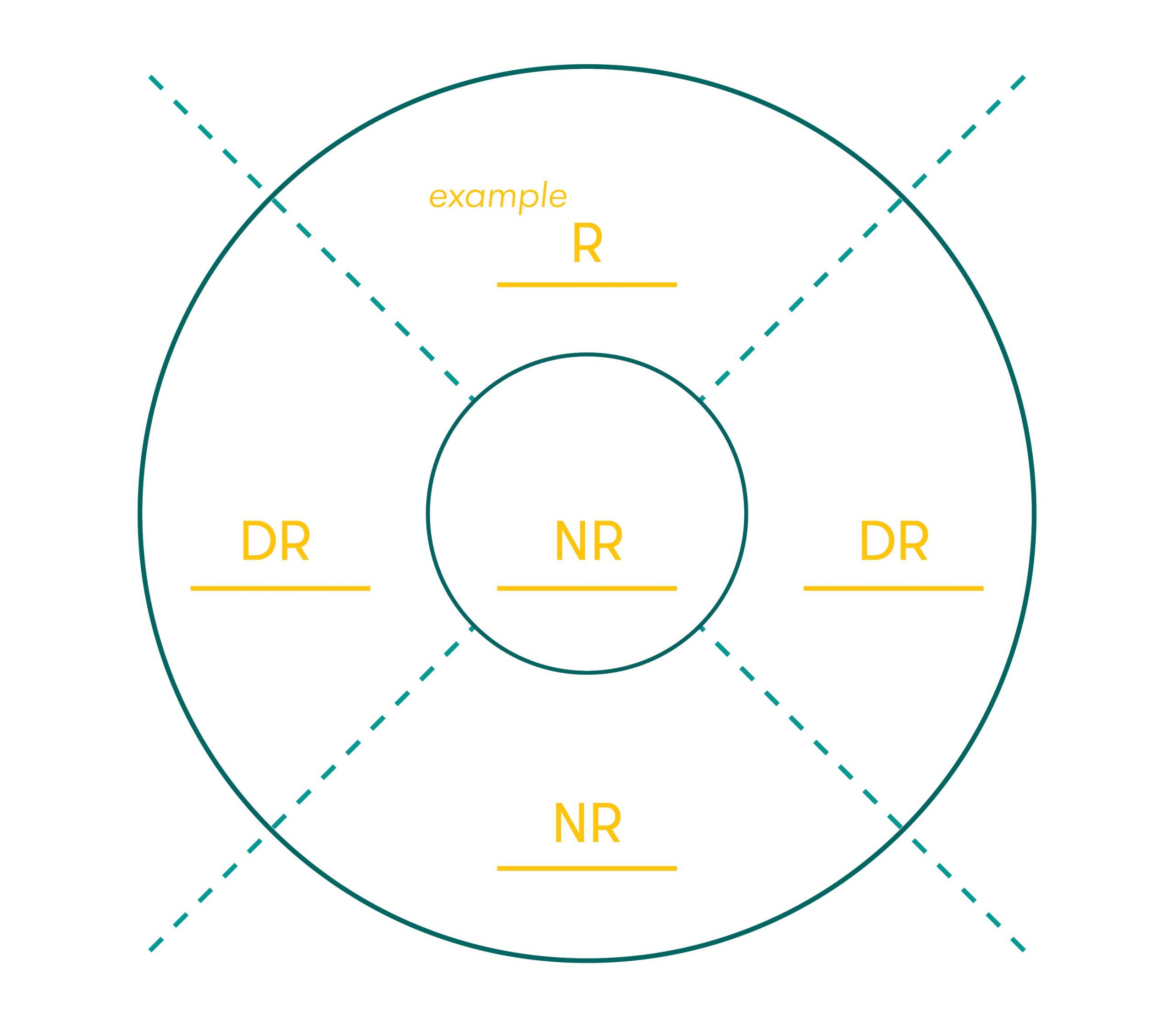
Assessment of corneal sensitivity is essential to confirm the diagnosis of NK and to assess the severity of corneal nerve impairment. Corneal sensitivity can be measured qualitatively by touching the central and peripheral cornea with a cotton thread or quantitatively using a corneal aesthesiometer.8 When the cotton thread gently touches the cornea, patients with a normal cornea show a blink reaction and can describe the sensation of touch, while patients with loss of corneal sensitivity do not react.

Label the chart with level of sensation using the Sensation Key.

Sensation Key

R – Responsive (Normal)

DR – Delayed Response (Reduced)

NR – Non-responsive (Absent)
Corneal Sensitivity Chart

As featured in Thomas John’s clinical study, there was a significant increase in corneal sensitivity from 1 to 3 months. This improvement was significantly correlated with the increase of corneal nerve density.7

The IVCM images showcase the subbasal nerve fiber and dendritiform cells in the study group baseline:

(a) 1 month

(b) 3 months

(c) at follow-up, thus proving there was a significant increase in corneal nerve regeneration.7


Download Dr. Thomas John’s Clinical Study on Corneal Nerve Regeneration.

By clicking the button below, I agree to receive marketing communications from BioTissue I am aware that I can unsubscribe at any time.
This field is for validation purposes and should be left unchanged.
References
Bonini S, Rama P, Olzi D, Lambiase A. Neurotrophic keratitis. Eye (Lond) 2003;17:989–995.
Bonini S, Lambiase A, Rama P, Sinigaglia F, Allegretti M, Chao W, Mantelli F, for the REPARO Study Group. Phase II randomized, double-masked, vehicle-controlled trial of recombinant human nerve growth factor for neurotrophic keratitis. Ophthalmology. 2018 Sep;125(9):1332-1343.
Faulkner WJ, Varley GA. Corneal diagnostic technique. In: Krachmer JH, Mannis MJ, Holland EJ, editors. Cornea: Fundamentals of Corneal and External Disease. St Louis, MO, USA: Mosby; 1997. 5. Dua HS, Said DG, Messmer EM, et al. Neurotrophic keratopathy. Prog Retin Eye Res. 2018;66:107-131.
Mead, O. G., Tighe, S., & Tseng, S. (2020). Amniotic membrane transplantation for managing dry eye and neurotrophic keratitis. Taiwan journal of ophthalmology, 10(1), 13–21. https://doi. org/10.4103/tjo.tjo_5_20
. Versura P, Giannaccare G, Pellegrini M, et al. Neurotrophic keratitis: current challenges and future prospects. Eye Brain. 2018 Jun 28;10:37-45.
John, T., Tighe, S., Sheha, H., Hamrah, P., Salem, Z. M., Cheng, A., ... & Rock, N. D. (2017). Corneal nerve regeneration after self-retained cryopreserved amniotic membrane in dry eye disease. Journal of ophthalmology, 2017.
Mead, O. G., Tighe, S., & Tseng, S. (2020). Amniotic membrane transplantation for managing dry eye and neurotrophic keratitis. Taiwan journal of ophthalmology, 10(1), 13–21. https://doi. org/10.4103/tjo.tjo_5_20
Kruse FE, Rohrschneider K, Völcker HE. Multilayer amniotic membrane transplantation for reconstruction of deep corneal ulcers. Ophthalmology. 1999;106:1504–10
Khokhar S, Natung T, Sony P, Sharma N, Agarwal N, Vajpayee RB. Amniotic membrane transplantation in refractory neurotrophic corneal ulcers: A randomized, controlled clinical trial. Cornea. 2005;24:654–60.
Nubile M, Dua HS, Lanzini M, Ciancaglini M, Calienno R, Said DG, et al. In vivo analysis of stromal integration of multilayer amniotic membrane transplantation in corneal ulcers. Am J Ophthalmol. 2011;151:809–220.
1. Mackie, I., Fraunfelder, F., & Roy, F. (1995). Current ocular therapy. Current Ocular Therapy. 4th ed. Philadelphia, PA: WB Saunders, 506-508